

SAN FRANCISCO, Aug. 29 (Xinhua) -- A new project, known as oVert, short for openVertebrate, will be launched in the United States to scan 20,000 vertebrates and make 3D images available online to researchers, educators, students and the public.
With a 2.5-million-dollar grant from the U.S. National Science Foundation (NSF), the project will enable researchers over four years to transport specimens from museum collections to computed tomography (CT) scanners, scan and upload images, and organize them on the public database MorphoSource for easy access.
While there are almost 70,000 vertebrate species described today, and more than half of those are fishes, the project will digitize more than one quarter of the world's vertebrate species, noted a recent news release from the University of Washington (UW), which will join 15 other institutions in the project.
"In a time when museums and schools are losing natural history collections and giving up due to costs, we are recognizing the information held in these specimens is only getting more valuable," project co-principal investigator Luke Tornabene, assistant professor of aquatic and fishery sciences at UW, was quoted as saying. "I think this project is going to help create a renaissance of the importance of natural history collections."
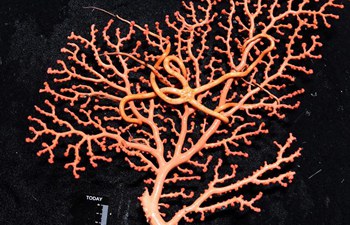
CT scanning is a non-destructive technology that bombards a specimen with X-rays from every angle, creating thousands of snapshots that a computer stitches together into a detailed 3D visual replica that can be virtually dissected, layer by layer, to expose cross-sections and internal structures. It allows scientists to view a specimen inside and out - its skeleton, muscles, internal organs, parasites, even its stomach contents - without touching a scalpel.
To be led by the Florida Museum of Natural History at the University of Florida, the team's largest scanner can image specimens as large as a garbage can, so for large mammals, researchers will focus on scanning their skulls or other key anatomical features; and some micro-CT scanners can pick up details of small vertebrates that are difficult to study at life size.
Tornabene, also curator of fishes at the Burke Museum of Natural History and Culture in Seattle, the largest city of Washington state in the U.S. Pacific Northwest, noted that UW researchers have already scanned some of the smallest fish in the world. In addition to being capable of zooming in to the digital file to examine anatomy not visible to the naked eye, they can 3-D print specimens larger than life.
"We are going to be exploring the capabilities of understanding vertebrate anatomy at the finest scales," he said.